DIRECT VISUALIZED RHIZOTOMY (DVR) SPINAL TREATMENT
Surgery is the last choice for treating back pain. Doctors and therapists usually try to treat chronic back pain with noninvasive methods first. However, back pain that lasts longer than 12 weeks is usually too severe for these kinds of treatment. In these cases, surgery is usually the best option for back pain treatment.
For years, spinal fusion and radiofrequency ablation have been the only proven options for the treatment of back pain that lasts longer than 12 weeks. These treatments can help people with spinal conditions reduce their pain, but it comes at a cost. Spinal fusion essentially joins separate vertebrae in your spine together, which results in the loss of mobility in your spine, is major surgery full of risks and often results in the need for additional surgery down the road. Radiofrequency ablation is not very invasive, but it’s often not effective, and when it does work the relief is temporary.
That’s why many people with chronic back pain that need something done are turning to direct visualized rhizotomy.
WHAT IS DIRECT VISUALIZED RHIZOTOMY?
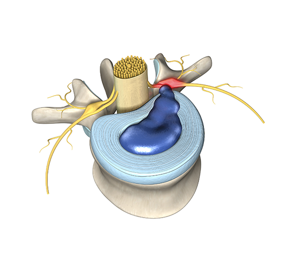
Rhizotomy is a treatment for chronic back pain that is an alternative to spinal fusion. The goal of rhizotomy is similar to the goals of a root canal. Rhizotomy treats back pain by cutting the ends of the pain fibers, which transmit the pain signal.
Rhizotomy can be taken a step further with the direct visualized rhizotomy procedure. Direct visualized rhizotomy can be more effective than radiofrequency ablation and much less invasive than spinal fusion. Direct visualized rhizotomy involves the use of an endoscope, which is a thin telescope with a camera on the end of it.
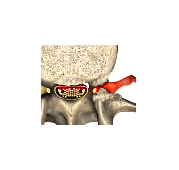
During your direct visualized rhizotomy procedure, your surgeon will insert an endoscopic tube through a very small incision (no bigger than five millimeters) and identify the medial branch of the spinal nerve root. Your surgeon will locate the branches of the nerve that are causing pain and sever them. After this, the endoscope is removed and the incisions are sealed with glue.
The procedure takes about 30 minutes and is usually completed under twilight sedation or general anesthesia if requested.
WHAT DOES DIRECT VISUALIZED RHIZOTOMY TREAT?
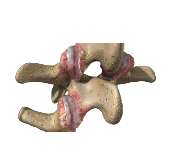
Direct visualized rhizotomy is used for back pain caused by facet joints. Facet joint syndrome, also referred to as spinal osteoarthritis, is a type of pain caused by the breaking down of cartilage in the joints of your spine. The pain typically begins on one side of your back, runs out into your buttocks, and feels like it’s in your hip. Severing the pain fibers with direct visualized rhizotomy can eliminate the pain that this condition causes.
Other conditions direct visualized rhizotomy can treat include:
- Ineffective radiofrequency ablation (RFA) treatments
- Sacroiliac joint disorder
- Back spasms caused by face joint problems
Your surgeon can assess your condition to determine if you’re the right candidate for direct visualized rhizotomy. There are three main indicators we check to determine if direct visualized rhizotomy will be right for you:
- They listen to your story to see if your pain fits the pattern caused by a swollen joint.
- The doctor reviews your MRI to confirm your joints are swollen and look for other causes of pain.
- The doctors perform a physical examination by pushing down on the precise area indicated by the MRI.
- They then numb the pain fibers that serve the joint. The numbing only lasts a few hours, but if it eliminates your pain during that time, it confirms the facet joint is the source of your pain.
WHAT ARE THE BENEFITS OF DIRECT VISUALIZED RHIZOTOMY?
Direct visualized rhizotomy is an ultra-minimally invasive procedure, which means there is little scarring and that you can recover quickly. Additionally, direct visualized rhizotomy preserves the integrity of your discs and vertebrae, which allows you to retain your freedom of mobility. Also, cutting the pain fiber is permanent; once cut, it can never transmit pain again.
Other benefits of direct visualized rhizotomy include:
- The procedure and its recovery time are fast
- The procedure does not involve cutting through your muscles or soft tissues
- Sedation is used instead of anesthesia in most cases
- Blood loss is minimal
- High success rates
- Permanent pain relief from the nerve fibers cut
- Less need for other treatments
ARE THERE ANY RISKS IN DIRECT VISUALIZED RHIZOTOMY?
Since the procedure is ultra-minimally invasive, the risks of infection are low. Look for facilities that have a zero percent infection rate.
It’s theoretically possible to cut the wrong nerve, but this is highly unlikely since direct visualized rhizotomy allows the doctor to visually identify the source of pain, which is more reliable than non-visual identification methods.
Some direct visualized rhizotomy patients are not good candidates for sedation, so they need anesthesia instead. Anesthesia is generally riskier than sedation, so there are slightly higher risks in these cases.
HOW LONG DOES IT TAKE TO RECOVER FROM DIRECT VISUALIZED RHIZOTOMY?
Patients are typically able to walk out of our facility under their own power within about an hour of the surgery’s completion. Some patients have felt so well after the procedure that they danced in celebration.
Many patients are able to return to work the next day. For some people, recovery may take a little longer, but this is okay. Your doctor will provide post-operative instructions to help you experience the best possible recovery.
PATIENT EDUCATION VIDEO
ADDITIONAL RESOURCES
International Journal of Medical Sciences Research on the use of DVR
Society for Minimally invasive Spine Surgery Study on the Long-Term Effects of Rhizotomy
A Study on DVR by International Journal of Spine Surgery
Learn about how we use a Medial Branch Block to diagnose not treat back pain


